Wednesday, December 12, 2012

It's narrow, regular, and fast - Now what?

Despite the variety of disorders an illness that parade through the ED, it often seems like each shift has a "theme." For example, you may diagnose two ectopic pregnancies in one shift, or send 3 patients to the cath lab. 

Heck, on a rare day you may actually have 3 or 4 patients with alcohol intoxication!

This is a thing that happens. Source

Last week for example, the theme for Yuko & I was narrow-complex tachycardias. As themes go, this was a good one - quick procedures, no untoward events, and we made the patients felt better!

Mrs Black
An older lady, Mrs Black had felt palpitations start about an hour before she arrived in the ED. Although she had no chest pain or pressure, she admitted to a little shortness of breath, as well as mild nausea. She took a variety of medications for hypertension, but no calcium-channel blockers or beta-blockers. Her vitals were normal.

Her first ECG:


Mrs. Black - before

After 6 mg of adenosine, we had:


Mrs. Black - after

Mr White
Our next PSVT came in only 2 hours later.

Also elderly, Mr White had been having some mild racing of his pulse over the past week, but the symptoms acutely worsened about 3 hours prior to coming to the ED. He had used a home-BP machine and noted that his systolic pressure had only been 70 at one point. The ED tech, of note, mentioned that he had looked fairly ill at triage - pale and sweaty.

His first ECG:


Mr White - before

And again, after 6 mg of adenosine (I'm not very original):


Mr White - after

So what's going on?
Ok, they're both narrow-complex tachycardias, very regular, with rates well under 150. So atrial fibrillation and flutter are pretty unlikely, as is a sinus tachycardia.

So that leaves the somewhat broad category of PSVT. Now, we usually just leave it at that in the ED, because, well, it doesn't usually matter to us whether the rhythm is caused by intranodal reentry (as in AV nodal reentrant tachycardia, or AVNRT) or extranodal reentry (as in AV reentrant tachycardia, or AVRT). This family of arrhythmias is almost always safe, even if the ECG shows ST depression, or we find a small troponin bump after conversion.

But the differentiation between different types of PSVTs is very important to the cardiologist, as there are implications for therapy. For example, AVRT is almost always primarily treated with an ablation. AVNRT, however, often is first treated a trial of medications (such as calcium-channel blockers, beta-blockers, or even digoxin), although ablation remains an option. 

So when one of the new cardiologists, Ram Gordon, from Cardiac Specialists, pointed out some interesting features on Mrs Black's ECG, we took the opportunity to expand our ECG skill set.


True fact: He's an ER fan!
Thanks to this brief, "just-in-time" teaching in the ED, when Mr. White was brought into the ER (just 2 hours after Mrs Black!), we were primed. Yuko & I only had to look at the ECG, and then at each other - we  had the diagnosis already.

Analyzing the initial ECGs
There's a good bit of cardiology literature devoted to making this diagnosis (AVRT vs AVNRT) using just the surface ECG. 

(If you want to do your own "deep dive" in this area, start of with these three articles, for example: Combined evaluation of bedside clinical variables and the electrocardiogram...Electrocardiographic differential diagnosis of narrow QRS complex tachycardia, and EGC diagnosis of paroxysmal supraventricular tachycardias...,.)

If, however, you don't feel like tackling the primary literature, you can instead check out this table, summarizing the elements that distinguish AVNRT form AVRT on the ECG:


Source: Gonzalez-Torrecilla 2011
Let's go use this information to evaluate the ECGs from Mr White and Mrs Black!

First let's look at Mrs Black's ECG. Are there indications of atrial activity? The best leads for this are usually II and V1. Typically the P-wave is upright in II, and inverted in V1, but a close look at those leads...
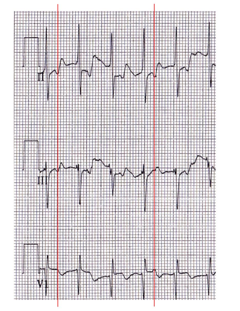
Detail of "Mrs Black - before"
... reveals inverted P's in II, falling just after the QRS complex - I'll say about 100 ms afterwards. It's also hard to ignore the ST depression in II and aVF!

These elements all suggest that this is an orthodromic AVRT, using a concealed pathway. Looks like the electrophysiologist has to go looking for a bypass tract!

Ok, what about Mr White?

You remember Mr White? He has a cool-sounding name.
A close look at Mr White's ECG shows...


Detail of "Mr White - Before"
... inverted P waves in II and III, with a suggestion of an upright P wave imposed on the T wave. Also, the RP interval is about 100 ms, and is distinct from the QRS complex. As long as that inverted P wave is distinct, and not "slurred" into a sort of "pseudo S' wave," it points to AVRT.

Pseudo S' in AVNRT, before & after conversion.   Source

So what?
So what's the big deal? Diagnosing an AVRT versus AVNRT in the ED isn't really crucial to the immediate management. So why should you try and delve deeper into the ECG? 

Well, first off, in EM we have the tough job of trying to sound smart to a lot of specialists (without ending up sounding too clever by half!). In any one shift, you'll find yourself describing an "overlapping distal radial fracture with intraarticular extension" to an orthopedist, a "perilimbal flush with consensual photophobia" to an ophthalmologist, and "He says he wants detox. Again." to the social worker. Similarly, in cardiology, we should using the language of their field, describing inverted Ps, pseudo S' waves, and such.

Furthermore, we can help point the patient in the right direction sooner. Evidence of AVRT, for example, suggests that we should talk with an electrophysiologist before discharge, arranging follow-up.


Lastly, you can't know too much about ECGs. Emergency physicians have to be experts in acute cardiology, give the pivotal role we have in the system. We can't afford to be second-best to anyone in the hospital, especially at 0300!

Thanks again to Dr Gordon for inspiring this post, and assisting with the cardiology perspective!

Wednesday, November 28, 2012

How to cure incomplete RBBB.

I had an interesting sign-out yesterday. The patient was a 30-ish year old woman with some vague complaint that localized to somewhere between her eyes and her knees. The off-going physician is a model of thoroughness, and they mentioned that the patient was getting, amongst other tests, a CT for pulmonary embolism.

"I wasn't really thinking PE at first," they explained to me, "but after I saw the incomplete RBBB on the ECG, I sent a D-dimer." The D-dimer, of course, came back trivially elevated, and the CT had been ordered.

Ah, the scourge of the IRBBB. It typically take the appearance of an rSR' pattern in V1 and V2 with a narrow QRS, although the AHA takes a dim view of this colloquial definition.

This seemingly "hard" element of evidence, however, can be surprisingly malleable!

I cured the patient's IRBBB!
The patient was already off at CT, so I looked at the ECG myself. It looked pretty darn normal, except for the right-precordial leads:


V2 shows an rSR' pattern, clearly.

The patient returned from CT scan, and I awaited the results. In the meantime, however, I re-interviewed an examined the patient, and convinced myself that she was low risk for all of the "bad actors." The IRBBB stuck in my craw, however, and after some fiddling around, I aquired a new ECG.

Sweet. It's not up there with sinking a difficult tube, or threading a pacer wire, but it makes for a cleaner chart!

What did I do?
I put V1 and V2 where they are supposed to go.

For a fuller explanation of the background here, please check out The most difficult step in obtaining an ECG. It's a post I wrote for paramedics about the importance, as well as the common difficulty, of placing the precordial leads in the correct locations. It's written in a conversational and witty tone, with plenty of illustrations - you'll like it!

Suffice it to say, though, that V1 and V2 are usually placed far too high on the chest. This produces a number of artifacts, including pseudo-infarction patterns and, yes, IRBBB.

I took a picture (with the patient's knowledge and permission - she believes in education!) of the actual lead placements on the patient's chest. The electrode wires are attached to the proper locations, at the 4th intercostal spaces, just to the right and left of the sternum.

Thank you Ms Anonymous!
The other electrode stickers are located only about 5-7 cm higher, around the 2nd or 3rd intercostal spaces, but it was enough to produce the spurious IRBBB.

The Bottom Line
Learn where to put the leads yourself.

If the diagnosis is going to rely critically on the ECG, you ought to check out where the electrodes were placed by the nurse, tech, or paramedic. Certainly, if there are abnormalities isolated to the precordial leads you should go back and do this, especially if the ECG doesn't match the patient's age or presentation.

BONUS QUESTION: For major extra points at the "Port, tell me what electrophysiologic diagnosis may rely on placing V1 and V2 higher than usual.

(Answer can be found by checking my self-aggrandizing link.)

*******
Added post-publication.

Mr Várhegyi points out a similar example, elsewhere on the web, of an IRBBB. Oddly, that example seemed to show some QRS prolongation in V2 that was not appreciated in the other leads.

Source

So, I took another look at my patient's initial ECG:


Darned if it doesn't seem to also have an isolated, minor (< 0.02 seconds), QRS prolongation in V2.

I'm going to defer providing an explanation until I have some idea about why this occurs, or even declaring that this is a reliable feature of improperly placed precordial leads. But it's apparent in these two examples. I'll look into it - anyone else with a better idea write in!


Monday, November 19, 2012

"Mystery" bradycardia, with some irony

Kito and I had an interesting patient last month. 

The patient
I had just been teaching a paramedic student some fine points of the ECG, when nurses asked us to come to room 41.

A 70 y.o. woman was brought in by EMS. She had syncoped when she stood up from the toilet. No zebras there - happens all the time! Her past medical history was about fair for her age; DM2, HTN, CAD, hyperlipidemia, etc, and on Plavix but no warfarin. She had gotten pretty banged up, with injuries to her face and leg.

What really stood out was that her heart rate; around 40. Her blood pressure was stable at 101/66, though, and she denied any chest pressure or trouble breathing.

The ECGs
Her first ECG was obtained at 18:24


A quick check of an old ECG (1 year prior) revealed that the LBBB, at least, was old:


Huh.

The Assessment
A review of her medications from her last ED visit listed carvedilol, and so accidental supratherapeutic dosing seemed likely. Line, labs, and imaging of her injuries were ordered, and we kept a close eye on the monitor.  Her blood pressure stayed north of 100, so we held off on pacers and pressors. Once she was cleared of any traumatic issues, she would be an easy CCU admit. Done.

Ah, not so fast...


 The Denouement

"This is the lab, and we have a critical value to report."

I actually like these phone calls - It's like a Maury Povich-type moment. You know that in the next few moments, all the pieces will fall into place, and you'll either be dancing around in self-congratulation, or applying both palms to your face firmly, rocking back and forth.


It wasn't the troponin, as I had been suspecting.

Instead, it was the potassium that turned up high - a level of 6.6 in fact. For some reason our patient, who had mild CKD before, now had a BUN of 104, and a GFR of 24.

Interestingly, renal and cardiology were not very impressed by the potassium level, feeling that it was a bit low to really cause much trouble. Kito & I had already started treatment, however. After 3 grams of calcium gluconate, the heart rate picked up, and we grabbed another ECG (21:17).


Seemed like we were on the right path, so we started dextrose and insulin, hung normal saline to address the AKI, and eventually gave some Kayexalate (Yes, I know what Weingart said on EmCrit, but a lot of folks still expect you to give it).

The Point

It's not just about tented T-waves.

I'm not going to review all of the protean ways that this electrolyte problem can manifest (please check out the comprehensive article by Matu et al).  I just want to emphasize how it showed up here.

First, there was an unexplained bradycardia, along with PR prolongation. Lastly, there was ST-segment depression in the anterior leads. In retrospect, these were classic signs of hyperkalemia, with plenty of examples from the literature.

Two cases, for example, describes complete AV block, but with a narrow QRS complex. Other cases demonstrate severe bradycardias without tented T-waves or pronounced QRS widening; e.g.

Case 1 from Atropine-resitant bradycardia due to hyperkalemia
You can even see some Wenkebach action, again without the dramatic T- or QRS-wave;

Wenckebach Block due to Hyperkalemia

You get the idea - you can go Google more examples if you want!

And the irony mentioned in the title?
As I mentioned above, I had been teaching ECG stuff to a medic student before this patient came in.  I could have been speaking with her about any number of topics - STEMIs, treating VT, recognizing atrial flutter... But no. 

Of course, it was about recognition of hyperkalemia on the ECG!


Saturday, November 3, 2012

STEMI with clean coronaries

The facts: a 35 year old male, with no medical history, presented with 1 week of chest pain that became acutely worse 1 hour prior. It was a "squeezing" feeling that radiated down his left arm. He had some mild dyspnea, and 1 nitro made it somewhat better. Some smoking, no cocaine.

The ECG:

The computer interpretation used CAPS LOCK, and had a lot of "***."

Cardiology was skeptical, but had him in the cath lab 30 minutes later. My resident put 50 cents down on a LAD occlusion, while I bet him a cup of (free) coffee that this was a classic first diagonal , or high lateral, STEMI. The two cardiology fellows agreed that we were both mistaken, and that they were certain to find a blocked circumflex. While the patient was in the lab the troponin came back as significantly elevated.

A few hours later, the cards fellow calls me back with the cath results.

Survey says!
No offense to Steve Harvey, but I'm a Dawson kinda guy.

Nada. Clean cath. "No significant fixed obstructive disease."

Interestingly, however, both ventriculography and an echo revealed hypokinesis of the high anterolateral wall, corresponding to the anatomy suggested by the ECG. He was given a diagnosis of focal myocarditis.

Focal Myocarditis
This isn't very common, but we can't say how uncommon. It is still uncommon enough to be worthy of case reports, at least in Texas. We know that about 3% of MIs (i.e. with positive cardiac enzymes) have clean coronary arteries by angiography, but a number of those people likely have spasm or spontaneous reperfusion. The percentage may even be smaller with true STEMI patterns, but we don't know.

The only way in the past to definitively diagnose myocarditis was through endomyocardial biopsy, which has a good number of shortcomings, both in terms of sensitivity, and of complications.

What could go wrong with this?
Advances in MRI techniques have enabled researchers to noninvasively study myocarditis. In a recent study it was found that 78% of patients who presented with an MI (64% with ST elevation), but a clean cath, had evidence of myocarditis on MRI.

Uh, yeah, I see it too...
Reciprocal changes
Now, I understand that the myocarditis can generate ST elevation, likely in the same manner that pericarditis does. I am really surprised, however, that our patient had such distinctive and appropriate reciprocal changes. Nonetheless, an ECG from a case report of myocarditis also shows reciprocal changes:


Turning to Stephan Smith's ECG Blog for some wisdom, I found this observation in "Is it MI or pericarditis?" (There's a lot of overlap between pericarditis and myocarditis, and many people link them on a spectrum; e.g myopericarditis.):
Pericarditis should never be assumed when there is even a hint of reciprocal ST depression.  Only localized pericarditis (most pericarditis is "diffuse" inflammation of the entire pericardium) ever has reciprocal ST depression, and localized pericarditis is very rare.  I suspect that many cases of "localized pericarditis" are really STEMI that went undiagnosed.

A great review article by Punja 2010 gives a few examples of ST elevation in myocarditis, but neither example shows reciprocal changes.

Nasty STE in myocarditis, but no ST depression
Sooo... Rare ECG finding? Not enough research? Incomplete diagnosis?

The Bottom Line

So, the next time you bring in that "for sure" STEMI, keep in mind there's a (3%*78%=) 2% chance it's myocarditis. Or higher. Or lower.

Thursday, July 12, 2012

Snuffbox tenderness: The D-dimer of orthopedics.

The conventional wisdom goes something like this:
Scaphoid fractures are a huge pitfall in emergency medicine. They can be subtle, and easily missed on the x-ray. If you find any tenderness in the anatomic snuffbox, you need to place a thumb spica splint, and have the patient referred to get a repeat x-ray in 7-10 days.
Bollocks. Pure felgercarb.

In a way, the EM approach to the suspected scaphoid fracture is a lot like our approach to the suspected PE. Based on data from the 60s and before, clinicians believe that a disease is difficult to clinically diagnose, radiographically elusive, and requires maximal therapy to avoid devastating outcomes. Some believe that new technologies and tests will provide an solution.

Maybe. But before we try to spend and irradiate our way out of this, let's take a step back, and look at some clinical wisdom you may not have heard about. Research that questions the "conventional wisdom."

And what better fount of conventional wisdom is there, than UpToDate? Let us examine some of the standard clinical advice from the article "Scaphoid Fractures."

Myth 1. Snuffbox tenderness is the cornerstone of diagnosis.
At Yale, there is no shortage of tweed-wearing attendings who solemnly intone rubbish about the decline of the physical exam, about the master diagnosticians of yesteryear, blah, blah, blah...

Cute. Now let's actually look inside.

Hey, you know why we use the ultrasound? Because the physical exams for AAA, CHF, peritoneal blood and gallstones aren't so good, that's why.

Uh, better stop tapping.
Many clinicians cannot recall any other test for scaphoid injury aside from snuffbox tenderness, and this isn't good, because the physical exam is key in determining your clinical suspicion here. This is also a problem since snuffbox tenderness is a lousy test, by itself.

UpToDate, however, assigns it a central role, despite having mentioned other exam techniques (my emphasis):
"Scaphoid fractures are often occult and a high index of suspicion should be maintained for any patient with wrist pain following trauma. Any tenderness in the snuffbox should be treated as a scaphoid fracture until proven otherwise."  (UTD)
This is unfortunate advice, since snuffbox tenderness, while usually very sensitive, is utterly non-specific. Parvizi 1998 found it to be only 19% specific, while Pillai 2005 only found 7% specificity!  Grover 1996 found 29%, and Freeland 1989 must have had a very light touch, determining a specificity of 40%. This makes the d-dimer look good.

With these shortcomings in mind, there are two other important exam techniques you should know:
  • Axial compression/impaction - Pressure is exerted in-line with the extended thumb, towards the radius. 
  • Tubercle tenderness - Palpate the bony prominence found on the distal flexor crease of the wrist. It is especially pronounced when the wrist is radially deviated and extended.
These aren't just "nice to know," or for historical interest. They might keep you from needlessly working patients up for fractures they don't have.

For instance, tubercle tenderness can be much more specific for fracture, but just as sensitive, according to Freeland 1989 and Parvizi 1998. Along the same lines, Grover 1996 found that the axial compression test was 80% specific, and 100% sensitive.

Parvizi 1998 found, in fact, that requiring a patient to be positive for all 3 exams would still be perfectly sensitive, while boosting specificity to 74%.

Myth 2. Despite negative intial x-rays, 10-25% of patients will have occult scaphoid fractures.
This actually phrased as
"[Splinting] of symptomatic patients with negative x-rays ... may result in approximately 75 to 90 percent of patients being immobilized for a week or more, [needlessly]." (UTD)
 Twenty five percent is a high number, and certainly would justify wrist immobilization if there's any doubt about the diagnosis. It would suggest that radiography is practically useless to detect fractures.

Except that Grover 1996 found that, of the 29 patients who had a scaphoid fracture in their series, all but one was picked up on the initial set of x-rays. Pillai 2005 only had a 7% rate of "occult" scaphoid fracture, while Duncan 1985 saw that none of their 108 patients had an "occult" fracture on follow-up.

So, your mileage may vary, but if both you and the radiologist don't see a fracture, you can feel pretty good about that.

Myth 3. If you don't immobilize the scaphoid soon, there is a huge risk of devastating complications.
The problem with a missed fracture is that non- or mal-union may cause the scaphoid to necrose, which leads to terrible functional outcomes. This is not rare, and it has lead to the perception that immobilization of the wrist needs to take precedence over all other considerations, such as cost, time off from work, and discomfort.
"[N]onunion rates can reach 40 percent when diagnosis and treatment are delayed by four weeks."(UTD)
The reference for this statement, Langhoff 1988, looked at a series of 289 scaphoid fractures. While there was an overall rate of non-union of 3-9%, they found no increase in this rate if the wrist was immobilized up to 28 days following the injury. On the other hand, after 28 days the rate of complications went up. The authors went on to conclude (my emphasis):
"As fractures not visible at the primary X-ray examination were identical with the rest as regards localization and healing characteristics, we assume that these fractures will behave in the same way when subjected to a delay of immobilization of less than four weeks. We therefore consider it unnecessary to immobilize the wrist when there is clinical suspicion of a fracture, but it is not demonstrable at X-ray examination."
They do consider it appropriate to obtain repeat x-rays at 2 weeks if clinical suspicion exists, and forgo immobilization until then.

Myth 4. The presumed scaphoid fracture must be immobilized in a thumb-spica splint.
Well, if my first 2 points are true, this is redundant. But for the sake of argument...
"When a definitive diagnosis cannot be determined at presentation and a scaphoid fracture is suspected on clinical grounds, even if radiographs are negative, the patient should be placed in a thumb spica splint or preferably, a thumb spica cast until a definitive imaging study can be performed." (UTD)
This is a pain in the ass to apply, and probably even worse to wear for the next 10 days, while you try to get into ortho clinic. Good luck using your hand for anything beyond a regal wave.

Hello!

Clay 1991 looked at patients with radiographic fractures, and randomized them to either a "Colles" cast, with the thumb waggling free, or a standard thumb spica.


The nonunion rate was the same in both groups.

Okay, maybe we don't need to immobilize the thumb. But surely we need a hard splint of some sort!

Eh, maybe not. Sjolin 1988 randomized patients with a clinical suspicion for scaphoid fracture, but negative x-rays,, to receive either a dorsal splint (no thumb immobilization) or a "supportive bandage" that could be removed for ADLs.

Everyone got rechecked in 2 weeks, and while there was no difference in complications (all fractures were incomplete or small avulsions), the bandage people were able to go back to work a lot sooner.

And residents need to be able to use their fingers!

Friday, July 6, 2012

Distinguishing STEMI from LVH on the ECG

I've started saying really something annoying at work.

When a resident starts interpreting an ECG, listing off features that either suggest or weigh against active ischemia, they will invariably (and properly) mention the deviation in millimeters of ST segments. I will then sanctamoniously intone

"Amateurs measure millimeters, professionals interpret the whole ECG."

If you think I'm smug about ECGs, ask about my thoughts on Huey Lewis.
Despite the my pretentiousness, there is an important element of truth there. (Dr Smith makes this point without smugness, but that's no fun. For me.) If you slavishly follow the standard criteria for STEMI diagnosis on the ECG (with so-and-so number of millimeters in this lead, so many leads with such elevation, etc.), you're going to make a lot of mistakes. 

One example is LBBB, a very common etiology of STE on the ECG. Usually, of course, the LBBB is immediately identified, owing to its characteristic appearance, and discussion switches to old vs new, and a review of Sgarbossa vs Smith criteria (peer-reviewed video lecture!).

The Problem with Left Ventricular Hypertrophy
Left ventricular hypertrophy (LVH), however, may not be as obvious, and ED physicians may mistakenly activate the cath lab based on the degree of ST segment elevation. Now, mistakes happen, and we want to have a certain "over-triage" rate, just like surgeons (before the use of CT scanners) had to have a certain "false appendectomy" rate. 

LVH, however, is very common, especially in the ED patient population. In fact, LVH is found in about a quarter of the ECGs in the ED that show STE, and is the most common cause.

Brady 2001
And it looks like emergency physicians get fooled by LVH fairly often. In one study of cath lab activations, it was found that LVH predicted a "false-postive" result, with an odds ration of 3.1.

How do we keep from getting fooled by this harmless (in the short-term) mimic of STEMI? It isn't simple. Indeed, one recent article suggested that "novice interpreters" of ECGs avoid diagnosing a STEMI in any patient with deep R- or S-waves.

Don't do it!

Well, I hope you are aspiring to more than "novice" status. I also hope that I can provide you some guidance beyond my usual semi-mystical injunctions and platitudes. 

"Be the ECG, Danny"
The Article


Fortunately, there are non-novice ECG interpreters out there who want to share their secrets, like this group out of UC-Davis. The authors used  a database ("ACTIVATE-SF) of all the emergency physician-initiated cath lab activations over a period of 3 years in the San Francisco area. This gave a denominator of 411 activations.

Applying any of 3 different scoring systems (Cornell, Sokolow-Lyon, or aVR > 11mm), they found that 79 of these patients met ECG criteria for LVH. Now, some of these patients with LVH ended up having culprit arteries identified at angiography, but most didn't. 

They went further, and analyzed this select group of patients who had LVH and a positive cath. They found a few predictors on the ECG.


Certain locations of STE, the degree of STE, and the number of leads with STE all increased the odds of a true STEMI. So did the presence of Q-waves or reciprocal ST depression.

Now, LVH usually only gives you a pseudo-STEMI appearance in the anterior leads, so elevations in the inferior leads, for example, won't cause much confusion. You can see from the table above that STE in the inferior or lateral leads, or a true posterior STEMI, will be easy to distinguish. But what we really care about is trying to distinguish an anterior MI from LVH, and the data above confirms the difficulty; STE in V1-V3 was more often found to not have a culprit artery.

ST Elevation: Absolute and Relative
 Rather than just measuring the absolute amount of STE, however, the authors also analyzed the relative amount of STE, normalized against the difference of the preceding R- and S-waves. 
% STE = (Height T-P segment to J-point) / (Height of R-wave minus depth of S-wave)
Here is an illustration of applying this, using an example of LVH without STEMI from the paper:


True STEMIs, overall, had a higher percent STE (25%) versus "false" STEMI (9%).  More importantly, if the percent STE < 25%, STEMI was essentially ruled out. If the percent STE > 25%, a STEMI was predicted with a sensitivity of 77%, and 91% specificity. 


When they compared this rule, using relative STE, versus the conventional criteria for STEMI that use absolute STE, they found it to be more sensitive and far more specific (73% and 58%, respectively).

Examples
For example this patient has LVH, and STE in V1-V3:


But they also have a ratio of STE/(R - S) = 45% STE


Indicating that it's a true STEMI.

Contrast this ECG with this patient with LVH:


They have STE, just like the preceding patient, but a closer look at the anterior leads shows...


... a fairly insignificant STE/R-S ratio, suggesting no active ischemia.

Putting it all together...
For practical use, the authors summarized, in an algorithm, the practical use of their findings, when interpreting an ECG with ST segment elevations.

The Bottom Line

Even if you don't crunch the numbers, try to appreciate the pattern, the proportionality, of the elevations and deviations. You need to look beyond the numbers, feel the waveforms, you need to...

Ah heck, I did it again.


Wednesday, March 28, 2012

The IMMEDIATE trial: Should EMS give Glucose-Insulin-Potassium?

The results of the IMMEDIATE trial have been popping up repeatedly today on Facebook, partly because I "like" a few EMS FB pages, and also because one of the authors (Hi Carin!) is a FB friend and recent Yale ED attending.

Here's an example of the way the trial is being described:


"ACS patients benefit;" "cut the risk of death in half." Sounds great! I love medical reporting/press releases. No pesky nuance or qualification. Me no need anyhow.

The result they are describing, to be specific, is that 8.7% of the people getting the placebo had a cardiac arrest, or died while they were hospitalized, while only 4.4% of the patients getting the study drug did. That's either an (absolute) difference of 4.3%, or about a (relative) 50% decline.

Such an effect would be stunning.  In the years after thrombolytics and aspirin were introduced, the incremental benefits of new therapies for AMI have been getting smaller and smaller. This result here would blow the others out of the water.

For instance, back in 1988, it was shown in ISIS-2 that either the use of aspirin or of thrombolytics reduced the risk of death in MI by about 2-3% over placebo. The combination was better of course.

After that, it's been harder to show that the more complicated and expensive therapies save that many more lives. When we send a patient to the cath lab for an AMI (instead of giving a thrombolytic in the ED), for example, there isn't that huge a benefit. One recent analysis suggested that, overall, you could only find a 0.7% difference in mortality (6.6% vs 5.9%) between lysed patients, and those that went for PCI. A lot of money for not much gain.

So, if this combination of glucose, insulin, and potassium (GIK) could cut mortality in AMI from 6.6% to, say, 3.3%, it would be freakin' amazing.

"I bet there's a catch. There's always a catch."
Well, I don't mean to be an Eeyore, but the perhaps we should wait for, yes, "further study." I offer three reasons why:

1. They weren't studying mortality.

The principle outcome they were studying was whether the initial presentation of ACS would progress to an MI, or it would be an "aborted" MI. This is the outcome that they believed had the most biochemical and clinical justification, and they clearly thought that it had a reasonable chance of being demonstrated.

It turns out there was no difference in the percent of people who progressed to completed MI - the GIK infusion did not help, at least not here. So the trial is negative for the real primary outcome.

2. There were 12 secondary outcomes.

Look at the table of the results:


Remember: the outcome they staked the success of the trial on was the one at the top: "Progression to MI," for all participants.  The rest are a bunch of secondary outcomes, and they don't count to the same degree as the primary outcome.

Analogy: A friend is flipping a coin, and you call heads. That's your primary outcome of interest. But if you also say to your friend "Okay, I call heads, but I also call it if you drop the coin, if it flips over 5 times in the air, if your phone rings in the next 30 seconds, or if your nose starts to itch in the next 10 seconds.

Now, you may be wrong about heads, but say your friend's nose does indeed start to itch in the next 10 seconds? Will he concede defeat? What will he say?

"No pick! NO PICK!" 
Most likely your friend will point out that the most relevant and important prediction you made was heads vs tails. Furthermore, you called out such a long list of other items that you were almost certain to come up with a positive result. He will insistent on another coin toss, where the primary outcome is now nose-itching, not heads or tails.

The same holds in statistics and study design, and is also why the authors state in their conclusion (my emphasis):
"The primary end point was not significantly different between groups, and the observed favorable results of GIK were based on prespecified but secondary end points, although biologically plausible and consistent with preclinical studies. The study tested one primary hypothesis, 3 major secondary, and 6 other secondary hypotheses. All were prespecified and no adjustment for multiple comparisons among the secondary end points was made; thus, reported significance levels should be considered approximate. Accordingly, given the lack of complete consistency of the findings, and the modest P values for most of the statistically significant findings, it would be appropriate to describe the observed favorable effects on the secondary outcomes as generating clinically testable hypotheses for evaluation in larger cohorts."

3. 30 day mortality seems pretty important too...

Ok, say you can take the "cardiac arrest or in-hospital mortality" results at face value. What, then, shall we make of the 30-day mortality? It was shown to be basically the same in both groups.

We just saw this discussion take place last month. A study from Japan showed that giving epinephrine in cardiac arrest got people to the hospital with ROSC more often, but the 30-day mortality was no different (We'll leave the neuro results alone for now.).

It would be nice if epi put all the dots on the right side of the graph. But it doesn't.
So, say the results are right - people don't die or arrest in the hospital as often, but they still die in the first 30 days just as often. Now, maybe everyone's hospital stay was over 30 days, but I doubt it.

Still feel excited?

Bottom line:

If they conduct another study that confirms the mortality benefit, it would be the greatest thing since the free coffee machine in the ED break room. But, unlike the coffee machine, such results are conjecture for now.