Sunday, November 13, 2011

Intraocular foreign bodies

Sometimes when I’m engaged in a “teaching moment” with a resident, a little voice will rouse itself and say “Hey! You know that last thing you said? It doesn’t sound quite right… You sure about that?” Sometimes it’s the resident saying this, of course (Looking at you, NewHavenResident!), but usually it’s my own inner voice.

I shared just such a moment with LeGrand a few days ago while we were discussing the possibility that a patient had an intraocular foreign body; the most common mechanisms, the clinical ophthalmologic exam, and the imaging test of choice. Somewhere in that conversation, I realized I was talking out of my hat. I decided a short blog post, going over some of the relevant literature, would be an appropriate penance!

First off, who gets these, and when does it happen?
Apparently, like most things in trauma, you are best off not being a young adult male. Three studies (in the UK, Egypt, and Malaysia) found that most injuries were incurred during work-related activities, and that firearms, explosions, and mowing the lawn can also play a large role.
  • Average patient is 29 – 38 y.o.
  • 92% - 100% are male.
  • In the developed world most IOFBs occur in the home (42%) rather than the workplace (33%).
  • Hammering still represents 60% to 80% of the cases.

    What kinds of things get shot into the eye? In one series of 74 IOFBs, 58 of these were metal, and 13 were glass, with only one wooden FB, and 2 “other.” A British series also found the vast majority were metal, with 9% being glass, stone, concrete, or, uh, eyelashes(?!).
    Eyelashes made of metal would explain a lot.
    The metal FBs can be drill bits, metal fillings from a lathe, as well as fragments of nails and other things you hit with a hammer. Various metals cause various problems – iron can stain the iris, copper causes a nasty inflammation, and lead can leech out sometimes.

    Other times, wood gets in the eye! Many of these FBs are from trees or branches, or are composed of some “treated” wood, which is consistent with occupational exposure. In one series, however, a third of wooden IOFBs were from pencils.
    Glass IOFBs, in one Indian series, was due to blast injury most often, as well as MVCs, and a smattering of other causes.
    Careful with your tumblers.

    What are you looking for on exam?
    So, how do you examine a patient when you have some suspicion for an IOFB? I’m not talking about the obvious cases, where you’re dialing for the on-call ophthalmologist right after you’re done with the A-B-Cs.
    "Uh, no, I haven't talked to his PMD yet..."
    Keep in mind that most IOFBs, especially glass shards, may be located in the posterior chamber, and may not be immediately obvious. A quick glance at the eyes to check off “PERRL” on the chart ain’t going to cut it – you need to bust out the ophthalmoscope, slit-lamp and fluoroscein.

    Key elements of the exam (Not exclusive).
                • Decreased visual acuity.
                • Deformation of the pupil.
                • Prolapsed iris.
                • Laceration of sclera, cornea.
                • Hyphema
                • Absence red reflex.
                • Seidel’s sign
    Click HERE to see the animation of a Seidel-positive exam. It's very cool!

    Now, if you see an FB at any point here, you’re done. Call optho, start some antibiotics, attend to the other injuries, etc. But if you haven’t found any direct evidence, you need to get some imaging. But what kind?

    Imaging for detection and characterization of an IOFB
    Plain films of the orbits are what the MRI techs ask for if there’s any history, even asymptomatic, of possible exposure to metal fragments in the past. But while they may have a place in screening low-risk patients for ferrous FBs, they don’t have much of a role in other patients.

    In one registry study, comprising both Hungary and U.S. patients, it was found that the clinical examination identified an IOFB in 46% of the patients examined, ultrasound revealed an IOFB 52% of the time it was used, and CT had 95% sensitivity when it was employed.

    A single-center study from a specialized eye & ear hospital in Ireland also looked at this question, conducting a chart review of patients with a suspicion of IOFB, who had had at least a plain film of the orbits. CT imaging of the orbits was only ordered if plain films had already been performed; only about 1 out of 10 patients went on to get a CT. Now, there was no gold standard here, and it seems like an IOFB was ruled out according to clinician judgment. Nonetheless, they found some encouraging results.
    • If there was no “clinically evident ocular penetration,” no IOFBs were found on plain film.
    • Similarly, no IOFBs were found on CT if the eye showed no clinical signs of penetration.
    • Interestingly, all patients who did have such signs either had a positive plain film or a positive CT scan.
    Given their results, they proposed a decision algorithm for imaging.

    Given the prime role that the clinical exam plays in their study results, as well as the algorithm, it is worth noting that the clinical exam these patients received went far beyond what many of us are comfortable with, including dilated fundoscopy and gonioscopy. Indeed, in the acutely injured patient, it may difficult to recognize an IOFB in the posterior chamber or elsewhere. With these factors in mind, the "EM-modified" algorithm boils down to…
    Very EM.
     Of course, in that study most of those FBs were metallic. When we look at imaging of non-metallic objects, the situation gets murkier.

    In one retrospective review of wooden IOFBs, CT was used in 22 out 23 cases. Even then, the radiologist could make a definitive call in less than 2/3 of the cases.
    "Cannot rule Ticonderoga #2. Clinical correlation suggested."
    In another study using an animal model of glass IOFBs, researchers found variable sensitivity for CT, MRI, and ultrasound in detection and identification. Interesting stuff: Detection rates were 57% for CT,  and 11% for T1-weighted MR. Ultrasound, meanwhile, found only 43% of glass fragments in the posterior chamber and 24% in the anterior chamber. On helical CT, anterior chamber glass was easiest to detect and corneal surface glass the most difficult. Sensitivity was greatest for green beer bottle glass (550 Hounsefield units!) and least for spectacle glass (around 80). So if you're going to be brawling at a saloon, take off your spectacles, and pick a beverage that comes in green glass!
    Or Heineken. Whichever.
    There are no good case series to look at the sensitivity of CT for other nonmetallic objects, but another animal model study may be helpful consider.  Researchers from Palermo used a pig model of the eye embedded with various materials, as well as injected air bubbles. The Italians found that the plain films were variable for detecting IOFBs, usually missing plastic or wooden objects. MRI was disappointing, showing significant artifact when examining objects made of  graphite, glass, and especially iron. The CT, however, was always able to “detect and differentiate” IOFBs.
    • Row A - Plain film
    • Row B - CT
    • Row C - MR-T1
    • Row D - MR-T2
    • Arrow = FB or air; arrowheads = lens; double arrows = optic nerve.

    But what about ultrasound?
    Ultrasound has some strong potential attributes. No radiation, no worries about jostling ferrous FBs around the head, and the patient isn't out of the department.

    And there are some encouraging studies to point to. One porcine model study found the technique to quite accurate, and found sensitivity to be 87%, and specificity 96%. However, the metallic fragments were introduced into the vitreous of the pig eye, whereas IOFBs "in real life" may not be as evident.

    This may be especially true with non-metallic objects. In the Indian study that surveyed their experience with glass IOFBs, the researchers noted that ultrasound could not detect the glass fragment in about 25% of patient. Typically the shards were located in either the anterior vitreous or ciliary body area. Artifacts from vitreous hemorrhage, lens opacities, or choroidal detachment also made for challenging ultrasound exams.

    A frequent concern about employing ultrasound, for whatever goal, is that it requires skill, and is (gasp!) operator-dependent. It was heartening to read one paper, then, that took on the issue of skill acquisition head-on. The authors of "Ultrasound detection of simulated intra-ocular foreign bodies by minimally trained personnel" wanted to see if they could teach 4 NASA astronauts to perform ocular ultrasound to a level comparable with a group of expert sonographers. They used a gelatinous ocular model embedded with various size pieces of metal, plastic, and glass.

    They are to be commended for taking the study to the obvious next level, and enrolled 10 high-school students in the study. I think you can guess where this is headed; the astronauts and high-school kids both got pretty good at picking out FBs, both groups demonstrating equal sensitivity and specificity, and not to far behind the experts!
    To be fair to John Glenn, they were from an AP biology class.
    Nonetheless, real-world use of ultrasound shows lower effectiveness. It's important to emphasize here that you should not be ultrasounding any eye that shows signs of a globe perforation, as the pressure of the probe could extrude contents. Furthermore, recall that a registry study mentioned earlier only found ultrasound to be 52% sensitive, far lower than CT. Now, ultrasound was only conducted when posterior chamber FBs or pathology was suspected, and likely was only employed if the object was not visualized on exam. Thus, the patients in whom ultrasound was the most appropriate were also patients in whom the exam would be more difficult!

    Take away message
    It may be surprising, after all that, to come to the nuanced conclusion:

    Just scan 'em.


    Thursday, November 3, 2011

    Benign early repolarization


    A few days ago I was just about to walk out of the pediatric ED, when the tech handed me an ECG: "I need you to look at this right now."


    All caps = important.
    All caps + asterisks = very important.


    Ah. I see. 
    The patient turned out to be a very healthy looking, comfortable, 28 y.o. African-American male, and he happened to have an odd chest discomfort, "like needles." He actually wasn't having the pain at the time the ECG was taken.

    Despite the dire diagnosis on the EKG (Thanks Marquette!), he walked out of the ED a few hours later, after multiple negative cardiac enzymes, sequential ECGs, a bedside echo, as well as a turkey sandwich. However, his favorable course could have been predicted from the classic findings on the ECG, diagnostic for benign early repolarization (BER).
    First off, before we review the criteria for BER, note that there are no reciprocal changes in the ECG, which would be extremely unlikely in a massive ***ACUTE MI***. Right off the bat, you know this is far more likely to represent a mimic of some ilk. 

    So, widespread ST elevations without reciprocal depression, there are 2 possibilities in the differential. Pericarditis is one, but a few things mitigate against it. There doesn't appear to any PR depression (or any PR elevation in aVR), and the T waves are all upright. So, possible, but not not clear.

    Now let's take a look at the criteria for BER.
    This table comes out of the excellent review article by Brady and Chan (1999) (Download pdf). Looking at the ECG, we see ST elevation in leads I, II, aVL, and V2-V5 - certainly very widespread, with elevations in the precordial leads looking more prominent than the limb leads. 

    Next, we note that in leads II and V5, where the ST elevation isn't that pronounced, there is nonetheless a notable elevation of the J-point, that place where the QRS "meets" the ST segment. 
    Lead II, showing J-point elevation over 0.1 mV
    Also note that none of the complexes with ST segment elevation show convex-upward segments, but instead concave-upwards morphology.

    Note also that the J-point in the ECG doesn't show a clean transition from the R wave to the ST segment. Rather, there seems to be a messy transition, either a "slurring" or a notched junction. You can see the slurred J-point in the blow-up from lead II above, or take a look at lead V4:
    Notched J-point
    As noted before, the T-waves are all appropriately upright and quite prominent. As for the other criteria, they're difficult to judge on a single ECG!

    Who has BER? Well, this patient was classic for the epidemiology of BER, being young, fit, male, and African-American.The pattern is found very often in male athletes, and appears to have no connection to HCM, or any other causes of sudden cardiac arrest.

    My philosophy is that the emergency physician needs to be competent in many spheres, and across many disciplines. However, in a few areas we need to be the experts in the hospital, and reading ECGs for signs of acute ischemia, or its mimics, is such a skill. In training we study more ECGS than anybody in any other field. And when you're an attneding in the ED, it's just one after another... All that training and repition pays off - In one study, Turnipseed et al. showed that EPs could read BER versus AMI on the ECG just as well as cardiologists, if they corrected for years of experience. (download pdf)

    See you in room 4!

    Tuesday, October 25, 2011

    Idiopathic Intercranial Hypertension; neither pseudo nor benign...

    Doug, sorry I had so few answers for you in the ED. Had to go hit the books after we had that patient!

    This isn't a comprehensive article on IIH, nor is it an efficient outline of the critical information for the boards, life, or pimping. It's just a list of 5 things that took me somewhat by surprise, that I had never learned, or that I swear I had learned as the opposite.

    1. "Transient visual obscurations" - The predominant visual disturbance isn't a nice, clean visual field cut. Instead, over 70% of patients have really brief episodes (< 1 minute) of loss of acuity, provoked by standing, sitting, light, Valsalva, or extra-ocular movements. Yeah, you'll get field loss on perimetry, but the TVOs are what patients will actually complain of.

    2. Sparkles - They can also get flashes, or photopsias, as well as the TVOs.They are described as "white or less commonly coloured small flashes, flickering lights, stars twinkling, or flashes in the periphery of the visual field
    Simulation available

    3. Patients with IIH also report an odd kind of noise inside their heads. It's termed "pulsatile tinnitus," but it only infrequently seems to be a high-pitched tone. More often, it's described as '"a rushing river', a waterfall or a buzzing, whistling or blowing sound."

    4. You get imaging to r/o something else, like a not-so-pseudo tumor or a sinus venous thrombosis. It's not used to diagnose IIH. However, one finding that would be consistent with IIH would be small ventricles or an empty sella,  according to some.
    Flattened ventricles.

    5. Perhaps the imaging study that we should be using more often is our ultrasound. Up until this month, the world's literature for using ultrasound to detect papilledema was limited to this study by Michael Stone (Download). While the case provided an example of using an optic nerve sheath diameter of >5 mm  to make a diagnosis of increased ICP, it also demonstrated sonographic papilledema.
    3mm posterior to the retina, check for OSD  > 5 mm.
    Also note optic disc elevation.


    But now there are 2 new publications that provide more evidence that we should be dropping our old clinical skills (polished as they may be...) in favor of using newer technology!
    1880 Loring opthalmoscope. Not new.
    The first article is an abstract that was just published in the October issue of Annals of Emergency Medicine, the ACEP Research Forum 2011 supplement, and is entitled "Point-of-Care Ocular Sonography to Detect Optic Disc Swelling." (Download pdf)

    The authors compared EP-performed ultrasound exams of the optic disc with those of an neuro-ophthalmologist, as well as optical coherence tomography.
    Optic disc swelling as seen on OCT
    For the study,  2 EPs hung out in a neuro-optho clinic, and did ocular US on a number of patients. They later compared their results with the specialist, who also had access to the OCT images and results. After 20 patients, they found they had "excellent correlation" between the OCT and height of the disc seen on ultrasound.
    Another example of an elevated optic disc (small arrow)
    Another article, still in-press at the American Journal of Emergency Medicine (by Daulaire et al.) (Download pdf), reviews a series of 3 headache patients who had apparent optic disc elevation on bedside US. The US results were considered confirmed, in each respective case, through a finding of elevated CSF pressure, an exam of the fundus by the ophthalmology service, and an MRI. There's a review of the technique, as well as of the scant literature.


    So, the next time you have that odd headache patient, ask them about TVOs and photopsias, and put a probe on their eyes!

    Saturday, October 22, 2011

    Cervicitis and PID

    It has come to my attention that the tenuous grasp I thought that I had possessed about cervicitis and PID became archaic at some point. My beloved, and much highlighted, text that was new when I started med school is now behind the times.
    It is so 2004...
    To be fair, the 2000's have been active in this area, and I think that you should hear a little bit about what's been goin' on. 

    First up: What are cervicitis and PID, i.e. what are the diagnostic criteria? This is important, because it turns out that in the ED we aren't so hot with the diagnosis of such things. Two studies point this out.
    That's pus.
    This study done in the Yale ED was written by your current boss (and with a future boss?), so you should probably know this! 
    Hint!
    The paper, "Compliance with the CDC Recommendations for the Diagnosis and Treatment of Sexually Transmitted Diseases" (Download here), published in 2004, documented how essential elements in the history and physical exam where missing from the charts of patients who had been diagnosed with cervicitis or PID. Furthermore, incomplete or erroneous treatment was often provided. All in an ED we all know and love...
    More pus in dere (PID).
    Another study, hot off the presses, make same case. (For download: Cervicitis in Adolescents: Do Clinicians Understand Diagnosis and Treatment?) Folks from the urology department at the U of Arkansas reviewed charts from both a primary care office and from a children's ED. All the records of adolescents who had received a diagnosis of cervicitis were analyzed for elements of the history, physical exam, and treatment. 

    First off, about 25% of the patients diagnosed with cervicitis did not have documented physical findings that supported the diagnosis; i.e. they had neither a mucopurulent cervical discharge, nor a friable, easily bleeding cervix. Despite this over-diagnosis, they also found that 40% of the women who had been diagnosed with cervicitis actually had sufficient exam finding to meet PID criteria. So, they were also under-treating! Urgh. 

    Even better, they break the results down according to the site of treatment: the adolescent clinic or the children's ED, and no one comes off looking good. The ED misdiagnosed about half the time, and the clinic about a third.

    So, let's get the definitions laid out, so there is no confusion. This all comes from the current CDC guide for STDs, which you can access online, or you can download the pdf.

    Cervicitis: 
    "Two major diagnostic signs characterize cervicitis: 1) a purulent or mucopurulent endocervical exudate visible in the endocervical canal or on an endocervical swab specimen (commonly referred to as mucopurulent cervicitis or cervicitis) and 2) sustained endocervical bleeding easily induced by gentle passage of a cotton swab through the cervical os." 

    PID:
    "Empiric treatment for PID should be initiated in sexually active young women and other women at risk for STDs if they are experiencing pelvic or lower abdominal pain, if no cause for the illness other than PID can be identified, and if one or more of the following minimum criteria are present on pelvic examination:
    • cervical motion tenderness
      or
    • uterine tenderness
      or
    • adnexal tenderness."
    They note that in cases where a diagnosis based on these minimal criteria "might cause unnecessary morbidity," that a few other elements may be used to boost the specificity, namely a mucopurulent discharge or a fever.
    Telling this guy that his 14 y.o. daughter has PID = morbidity?
    By contrast, the 1998 CDC guidelines required that all 3 of the minimal criteria (CMT, uterine, and adnexal tenderness) be found in order to make a diagnosis. Why did this change? After all, it's not so often that we change a diagnosis. Femur fractures, for example, have never had a revision of the diagnostic criteria!

    Well, the PEACH trial happened. (Download here
    This was a major randomized trial, and I simply will not do justice summarizing it here. Instead of reading any more mind-numbing studies about subarachnoids or PEs, read this. 

    The trail was designed to examine the effectiveness of IV versus outpatient treatment of PID, but since they were collecting all this data on women with suspected PID, and then getting all these confirmatory tests, they wanted to see how sensitive and specific the elements of the CDC diagnostic criteria were. It was a multicenter trial, in both clinics and EDs throughout the US. Diagnostic techniques were comprehensive: PID was diagnosed histologically, from samples obtained from transcervical aspiration of endometrial tissue. In the end, they had complete information on 651 women, and the results were...
    They found that the minimal CDC criteria (all 3 of CMT, adnexal, and uterine tenderness) was only 83% sensitive, with a specificity that wasn't exactly a selling point. Furthermore, if you were hoping that supporting elements, such as a fever (or it's absence) might help diagnosis, there were disappointing results.
    For my part, the negative likelihood ratios for fever and purulent discharge grab my attention. The absence of the elements does not meaningfully help the diagnosis, since the negative LR is close to 1. Heck, even a negative NAAT test only cuts the probability in half!

    In the discussion section, the authors offered their suggestion that "clinicians should consider empiric treatment of pelvic inflammatory disease in at-risk women with adnexal tenderness at presentation and no other obvious diagnosis," which is close to the current guidelines. And even with this incredibly liberal definition of PID, we are still going to miss about 5%!

    So that's it for diagnosis. As for treatment, like all of ID, things change every year - look up the current reccs in the CDC guide I linked above. Some things are suprising (no more 125 of Rocephin!), and others stay the same (7 days of doxy for cervicitis, 14 for PID).This post is long enough, so I'll leave you with just one more cervix:
    Bye!

    Thursday, October 6, 2011

    2 important points about treating ACS

    I wanted to quickly review 2 issues that come up with regularity when a resident & I have a patient with ACS, either a STEMI or NSTEMI. Let me start with the conclusion first, and go from there.

    1. Tachycardia is a reason not to give IV beta-blockers in the ED for NSTEMI or STEMI.
    2. Heparin hasn't actually been shown to have much effect in NSTEMI.

    Okay, first about the beta-blockers. There used to be a lot more enthusiasm for using IV metoprolol in the ED with a STEMI, and many clinicians still feel that they are obligated to give them. But a lot of that changed, first when the COMMIT trial came out, and then when the AHA modified their guidelines to reflect the new evidence. In that big trial, patients with a STEMI were given the IV metoprolol load in the ED, followed by PO. The kicker is that there was overall no difference in 28 day mortality, since for every person who apparently didn't have a reinfarct or fatal arrhythmia. there was an extra person who died of HF!

    Make special note of the risk factors listed after the asterix, in small print, at the bottom: sinus tachycardia exceeding 110 bpm.

    These considerations were extended to the NSTEMI guidelines as well, although the evidence base is not as direct or contemporary as it is for STEMI. The 2007 AHA UA/NSTEMI guidelines basically mirror those for STEMI with regard to PO beta-blockers (Class 1 in the first 24 hours) and IV beta-blockers (class 2a, with same contraindications as above).


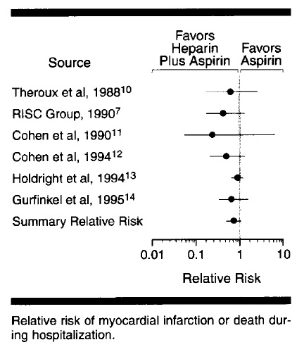
    As for heparin for NSTEMI, it is sort of absurd looking at the recommendations and the supporting evidence. The 2007 AHA UA/NSTEMI guidelines cite 2 studies in support of the use of UFH. The more recent, and larger, was  this paper , a meta-analysis of 6 studies that looked at the benefit of adding heparin to aspirin in UA/NSTEMI.
     Ok, look at the title. Seems like a pretty clear title - you know just what they found in their study, and you should expect to find at least statistical significance in the results, right?
    Not really
    Right there, first page - the RR for MI or death was not significantly reduced when heparin was added to aspirin. They include this helpful graph, which shows the results of 6 trials, and the summary estimate that they calculate.
    In the last few sentences, the authors, note that "this meta-analysis of 6 randomized controlled trials demonstrated a strong trend toward reduction in risk of MI or death during randomized therapy in patients with unstable angina treated with aspirin plus heparin compared with those treated with aspirin alone." The phrase "strong trend" is not found in the conclusion of the abstract, but instead they highlight the "33% reduction" in outcomes. 

     Heparin probably has utility in the sicker subset of NSTEMI, the patients with positive enzymes, dynamic ECG changes, ongoing pain. But those are the kind of patients you're talking about with the cardiologist, calling CCU, and perhaps arranging same-day cath lab evaluation. But for the run o' the mill ACS patient, check out the evidence first.

    Tuesday, September 20, 2011

    Resources for Acute Stroke evaluation and treatment

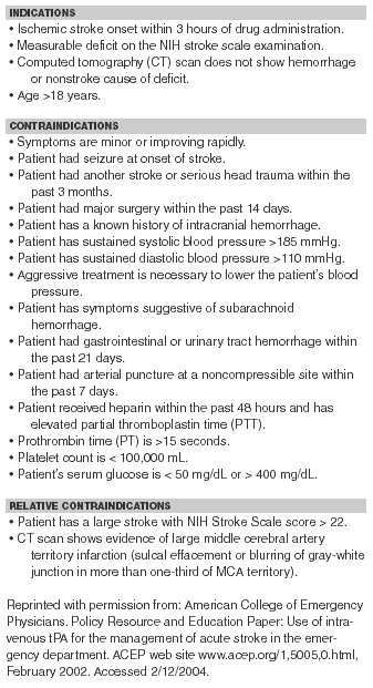
    This post is really just for me - so I can find a few certain documents without frantic Googling when the 3 hour clock is ticking...

    AHA/ASA - Guidelines for the Early Management of Adults With Ischemic Stroke

     ACEP - Use of IV tPA for the Management of Acute Stroke in the ED


    AHA/ASA Science Advisory - Expansion of the Time Window for Treatment of Acute Ischemic Stroke With Intravenous Tissue Plasminogen Activator (del Zoppo)

    ECASS III Exclusion Criteria 
    http://crashingpatient.com/medicine-surgery/095-stroke.htm  
    Expansion of TPA window to 4.5 hours by AHA (Stroke 2009;40:) - ECASS III
    • Age < 18 or > 80 years
    • Onset of stroke > 4.5 hours before drug administration or symptom onset unknown
    • Stroke symptoms present < 30 minutes or significantly improving before treatment
    • Intracranial hemorrhage
    • Severe stroke as defined by NIHSS > 25 or imaging (CT or MRI) displaying > 1/3 of middle cerebral artery territory involved
    • Seizure at the onset of stroke
    • --------
    • Stroke or serious head trauma within the previous 3-months 
    • Major surgery or severe trauma within 3-months
    • Combination of previous stroke and diabetes mellitus
    • --------
    • SBP > 185 mm Hg or DBP > 110 mm Hg or IV treatment to reduce BP to these limits
    • Glucose < 50 mg/dL or > 400 mg/dL
    • Symptoms suggestive of subarachnoid hemorrhage even if CT normal
    •  -------
    • Heparin within the preceding 48 hours with PTT above normal limit
    • Platelet count < 100,000 mm3
    • Oral anticoagulation therapy (EVEN IF INR WNL)
    • Other major disorders with an increased risk of bleeding


    www.aaem.org/education/tpaedtool-AAEM.pdf

    Monday, September 19, 2011

    Transfusion triggers

    Quick note today, just wanted to talk about the hemoglobin level at which we should consider transfusion.

    There's a lot of bad data out there, much of it retrospective, heterogeneous populations, and so on. When I was a MICU resident, often the team would prefer to get the Hgb up above 10 to "help with the oxygen delivery." Marino, OTOH, in his The ICU Book, makes a persuasive case that the reduced viscosity in anemia facilitates oxygen delivery, and you mess with this compensatory mechanism (with pRBCs) at your peril. Incidently, Marino provides a review of some more recent transfusion-related papers from his website. Worth reading.

    Let me review the single best, and most-oft cited, paper on this topic, which was done by Hêbert et al, published in the NEJM in 1999. The paper (pdf) can be downloaded free from NEJM, I think, but here's a copy.
    Real interesting stuff.

    They prospectively studied the effect of two different transfusion thresholds, either 7 g/dL (restrictive strategy) or 10 g/dL (liberal strategy), in ICU patients. The ICUs were mixed, both surgical and medical, and both community and academic. They excluded patients with, amongst other issues,  active hemorrhage or s/p cardiac surgery.
    They found that 30-day mortality was the same in the two groups, but the hospitalization mortality was significantly lower in the restrictive-strategy group (22.3 percent vs. 28.1 percent, P=0.05).

    Let me emphasize that a bit more:
    28.1 - 22.3 = 5.8,  or an Absolute Rate Reduction of 5.8%
    That gives us a Number Needed to Harm of a little over 17. In other words:

    For every 17 patients that were transfused to stay above 10 g/dL, instead of above 7 g/dL, 1 died.


    Closer look at the breakdown on adverse effects:
    Hey, that's funny. Many folks feel that a chief reason to transfuse is to avoid exacerbating any cardiac ischemia. What they found here, however, was that bad cardiac events were more common with the liberal transfusion threshold!

    Okay, that was in 1999 - what's changed since then? Not much. In fact, the evidence has become stronger with regard to the risks of transfusion, and the benefits of a restrictive strategy, across a spectrum of medical and surgical contexts.

    Let me give the latest transfusion guidelines, published in 2009, written jointly by the Society for Critical Care Medicine,and the Eastern Association for Surgery on Trauma. This document is kinda unique, addressing both the medical and surgical perspectives regarding the literature. The salient recommendations are copied here. I just want to highlight a few of them:

    1. Transfuse patients with active, ongoing hemorrhage. Be proactive!
    2. If they're not bleeding out, don't transfuse until the Hgb drops below 7 g/dL.
    3. At that point, only transfuse single units.

      A. Recommendations Regarding Indications for RBC Transfusion in the General Critically Ill Patient
        1.    RBC transfusion is indicated for patients with evidence of hemorrhagic shock. (Level 1)  
        2.    RBC transfusion may be indicated for patients with evidence of acute hemorrhage and hemodynamic instability or inadequate oxygen delivery. (Level 1)  
        3.    A “restrictive” strategy of RBC transfusion (transfuse when Hb < 7 g/dL) is as effective as a “liberal” transfusion strategy (transfusion when Hb < 10 g/dL) in critically ill patients with hemodynamically stable anemia, except possibly in patients with acute myocardial ischemia. (Level 1)  
        4.    The use of only Hb level as a “trigger” for transfusion should be avoided. Decision for RBC transfusion should be based on an individual patient's intravascular volume status, evidence of shock, duration and extent of anemia, and cardiopulmonary physiologic parameters. (Level 2) 
        5.    In the absence of acute hemorrhage RBC, transfusion should be given as single units. (Level 2)  
        6.    Consider transfusion if Hb < 7 g/dL in critically ill patients requiring mechanical ventilation (MV). There is no benefit of a “liberal” transfusion strategy (transfusion when Hb < 10 g/dL) in critically ill patients requiring MV. (Level 2)  
        7.    Consider transfusion if Hb < 7 g/dL in resuscitated critically ill trauma patients. There is no benefit of a “liberal” transfusion strategy (transfusion when Hb < 10 g/dL) in resuscitated critically ill trauma patients. (Level 2)  
        8.    Consider transfusion if Hb < 7 g/dL in critically ill patients with stable cardiac disease. There is no benefit of a “liberal” transfusion strategy (transfusion when Hb < 10 g/dL) in critically ill patients with stable cardiac disease. (Level 2) 
        9.    RBC transfusion should not be considered as an absolute method to improve tissue oxygen consumption in critically ill patients. (Level 2) 
        10.  RBC transfusion may be beneficial in patients with acute coronary syndromes (ACS) who are anemic (Hb ≤ 8 g/dL) on hospital admission. (Level 3)
    ...