Tuesday, October 25, 2011

Idiopathic Intercranial Hypertension; neither pseudo nor benign...

Doug, sorry I had so few answers for you in the ED. Had to go hit the books after we had that patient!

This isn't a comprehensive article on IIH, nor is it an efficient outline of the critical information for the boards, life, or pimping. It's just a list of 5 things that took me somewhat by surprise, that I had never learned, or that I swear I had learned as the opposite.

1. "Transient visual obscurations" - The predominant visual disturbance isn't a nice, clean visual field cut. Instead, over 70% of patients have really brief episodes (< 1 minute) of loss of acuity, provoked by standing, sitting, light, Valsalva, or extra-ocular movements. Yeah, you'll get field loss on perimetry, but the TVOs are what patients will actually complain of.

2. Sparkles - They can also get flashes, or photopsias, as well as the TVOs.They are described as "white or less commonly coloured small flashes, flickering lights, stars twinkling, or flashes in the periphery of the visual field
Simulation available

3. Patients with IIH also report an odd kind of noise inside their heads. It's termed "pulsatile tinnitus," but it only infrequently seems to be a high-pitched tone. More often, it's described as '"a rushing river', a waterfall or a buzzing, whistling or blowing sound."

4. You get imaging to r/o something else, like a not-so-pseudo tumor or a sinus venous thrombosis. It's not used to diagnose IIH. However, one finding that would be consistent with IIH would be small ventricles or an empty sella,  according to some.
Flattened ventricles.

5. Perhaps the imaging study that we should be using more often is our ultrasound. Up until this month, the world's literature for using ultrasound to detect papilledema was limited to this study by Michael Stone (Download). While the case provided an example of using an optic nerve sheath diameter of >5 mm  to make a diagnosis of increased ICP, it also demonstrated sonographic papilledema.
3mm posterior to the retina, check for OSD  > 5 mm.
Also note optic disc elevation.


But now there are 2 new publications that provide more evidence that we should be dropping our old clinical skills (polished as they may be...) in favor of using newer technology!
1880 Loring opthalmoscope. Not new.
The first article is an abstract that was just published in the October issue of Annals of Emergency Medicine, the ACEP Research Forum 2011 supplement, and is entitled "Point-of-Care Ocular Sonography to Detect Optic Disc Swelling." (Download pdf)

The authors compared EP-performed ultrasound exams of the optic disc with those of an neuro-ophthalmologist, as well as optical coherence tomography.
Optic disc swelling as seen on OCT
For the study,  2 EPs hung out in a neuro-optho clinic, and did ocular US on a number of patients. They later compared their results with the specialist, who also had access to the OCT images and results. After 20 patients, they found they had "excellent correlation" between the OCT and height of the disc seen on ultrasound.
Another example of an elevated optic disc (small arrow)
Another article, still in-press at the American Journal of Emergency Medicine (by Daulaire et al.) (Download pdf), reviews a series of 3 headache patients who had apparent optic disc elevation on bedside US. The US results were considered confirmed, in each respective case, through a finding of elevated CSF pressure, an exam of the fundus by the ophthalmology service, and an MRI. There's a review of the technique, as well as of the scant literature.


So, the next time you have that odd headache patient, ask them about TVOs and photopsias, and put a probe on their eyes!

Saturday, October 22, 2011

Cervicitis and PID

It has come to my attention that the tenuous grasp I thought that I had possessed about cervicitis and PID became archaic at some point. My beloved, and much highlighted, text that was new when I started med school is now behind the times.
It is so 2004...
To be fair, the 2000's have been active in this area, and I think that you should hear a little bit about what's been goin' on. 

First up: What are cervicitis and PID, i.e. what are the diagnostic criteria? This is important, because it turns out that in the ED we aren't so hot with the diagnosis of such things. Two studies point this out.
That's pus.
This study done in the Yale ED was written by your current boss (and with a future boss?), so you should probably know this! 
Hint!
The paper, "Compliance with the CDC Recommendations for the Diagnosis and Treatment of Sexually Transmitted Diseases" (Download here), published in 2004, documented how essential elements in the history and physical exam where missing from the charts of patients who had been diagnosed with cervicitis or PID. Furthermore, incomplete or erroneous treatment was often provided. All in an ED we all know and love...
More pus in dere (PID).
Another study, hot off the presses, make same case. (For download: Cervicitis in Adolescents: Do Clinicians Understand Diagnosis and Treatment?) Folks from the urology department at the U of Arkansas reviewed charts from both a primary care office and from a children's ED. All the records of adolescents who had received a diagnosis of cervicitis were analyzed for elements of the history, physical exam, and treatment. 

First off, about 25% of the patients diagnosed with cervicitis did not have documented physical findings that supported the diagnosis; i.e. they had neither a mucopurulent cervical discharge, nor a friable, easily bleeding cervix. Despite this over-diagnosis, they also found that 40% of the women who had been diagnosed with cervicitis actually had sufficient exam finding to meet PID criteria. So, they were also under-treating! Urgh. 

Even better, they break the results down according to the site of treatment: the adolescent clinic or the children's ED, and no one comes off looking good. The ED misdiagnosed about half the time, and the clinic about a third.

So, let's get the definitions laid out, so there is no confusion. This all comes from the current CDC guide for STDs, which you can access online, or you can download the pdf.

Cervicitis: 
"Two major diagnostic signs characterize cervicitis: 1) a purulent or mucopurulent endocervical exudate visible in the endocervical canal or on an endocervical swab specimen (commonly referred to as mucopurulent cervicitis or cervicitis) and 2) sustained endocervical bleeding easily induced by gentle passage of a cotton swab through the cervical os." 

PID:
"Empiric treatment for PID should be initiated in sexually active young women and other women at risk for STDs if they are experiencing pelvic or lower abdominal pain, if no cause for the illness other than PID can be identified, and if one or more of the following minimum criteria are present on pelvic examination:
  • cervical motion tenderness
    or
  • uterine tenderness
    or
  • adnexal tenderness."
They note that in cases where a diagnosis based on these minimal criteria "might cause unnecessary morbidity," that a few other elements may be used to boost the specificity, namely a mucopurulent discharge or a fever.
Telling this guy that his 14 y.o. daughter has PID = morbidity?
By contrast, the 1998 CDC guidelines required that all 3 of the minimal criteria (CMT, uterine, and adnexal tenderness) be found in order to make a diagnosis. Why did this change? After all, it's not so often that we change a diagnosis. Femur fractures, for example, have never had a revision of the diagnostic criteria!

Well, the PEACH trial happened. (Download here
This was a major randomized trial, and I simply will not do justice summarizing it here. Instead of reading any more mind-numbing studies about subarachnoids or PEs, read this. 

The trail was designed to examine the effectiveness of IV versus outpatient treatment of PID, but since they were collecting all this data on women with suspected PID, and then getting all these confirmatory tests, they wanted to see how sensitive and specific the elements of the CDC diagnostic criteria were. It was a multicenter trial, in both clinics and EDs throughout the US. Diagnostic techniques were comprehensive: PID was diagnosed histologically, from samples obtained from transcervical aspiration of endometrial tissue. In the end, they had complete information on 651 women, and the results were...
They found that the minimal CDC criteria (all 3 of CMT, adnexal, and uterine tenderness) was only 83% sensitive, with a specificity that wasn't exactly a selling point. Furthermore, if you were hoping that supporting elements, such as a fever (or it's absence) might help diagnosis, there were disappointing results.
For my part, the negative likelihood ratios for fever and purulent discharge grab my attention. The absence of the elements does not meaningfully help the diagnosis, since the negative LR is close to 1. Heck, even a negative NAAT test only cuts the probability in half!

In the discussion section, the authors offered their suggestion that "clinicians should consider empiric treatment of pelvic inflammatory disease in at-risk women with adnexal tenderness at presentation and no other obvious diagnosis," which is close to the current guidelines. And even with this incredibly liberal definition of PID, we are still going to miss about 5%!

So that's it for diagnosis. As for treatment, like all of ID, things change every year - look up the current reccs in the CDC guide I linked above. Some things are suprising (no more 125 of Rocephin!), and others stay the same (7 days of doxy for cervicitis, 14 for PID).This post is long enough, so I'll leave you with just one more cervix:
Bye!

Thursday, October 6, 2011

2 important points about treating ACS

I wanted to quickly review 2 issues that come up with regularity when a resident & I have a patient with ACS, either a STEMI or NSTEMI. Let me start with the conclusion first, and go from there.

1. Tachycardia is a reason not to give IV beta-blockers in the ED for NSTEMI or STEMI.
2. Heparin hasn't actually been shown to have much effect in NSTEMI.

Okay, first about the beta-blockers. There used to be a lot more enthusiasm for using IV metoprolol in the ED with a STEMI, and many clinicians still feel that they are obligated to give them. But a lot of that changed, first when the COMMIT trial came out, and then when the AHA modified their guidelines to reflect the new evidence. In that big trial, patients with a STEMI were given the IV metoprolol load in the ED, followed by PO. The kicker is that there was overall no difference in 28 day mortality, since for every person who apparently didn't have a reinfarct or fatal arrhythmia. there was an extra person who died of HF!

Make special note of the risk factors listed after the asterix, in small print, at the bottom: sinus tachycardia exceeding 110 bpm.

These considerations were extended to the NSTEMI guidelines as well, although the evidence base is not as direct or contemporary as it is for STEMI. The 2007 AHA UA/NSTEMI guidelines basically mirror those for STEMI with regard to PO beta-blockers (Class 1 in the first 24 hours) and IV beta-blockers (class 2a, with same contraindications as above).


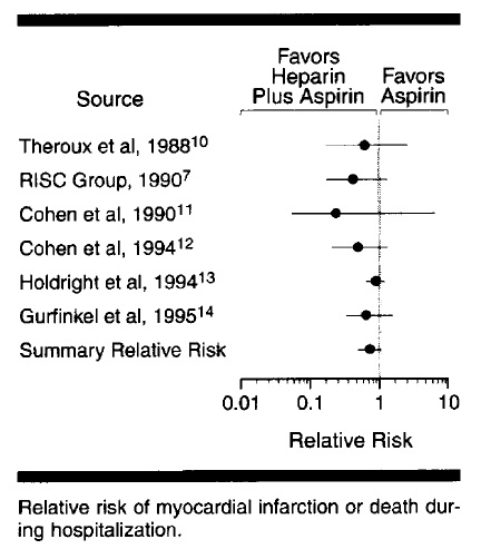
As for heparin for NSTEMI, it is sort of absurd looking at the recommendations and the supporting evidence. The 2007 AHA UA/NSTEMI guidelines cite 2 studies in support of the use of UFH. The more recent, and larger, was  this paper , a meta-analysis of 6 studies that looked at the benefit of adding heparin to aspirin in UA/NSTEMI.
 Ok, look at the title. Seems like a pretty clear title - you know just what they found in their study, and you should expect to find at least statistical significance in the results, right?
Not really
Right there, first page - the RR for MI or death was not significantly reduced when heparin was added to aspirin. They include this helpful graph, which shows the results of 6 trials, and the summary estimate that they calculate.
In the last few sentences, the authors, note that "this meta-analysis of 6 randomized controlled trials demonstrated a strong trend toward reduction in risk of MI or death during randomized therapy in patients with unstable angina treated with aspirin plus heparin compared with those treated with aspirin alone." The phrase "strong trend" is not found in the conclusion of the abstract, but instead they highlight the "33% reduction" in outcomes. 

 Heparin probably has utility in the sicker subset of NSTEMI, the patients with positive enzymes, dynamic ECG changes, ongoing pain. But those are the kind of patients you're talking about with the cardiologist, calling CCU, and perhaps arranging same-day cath lab evaluation. But for the run o' the mill ACS patient, check out the evidence first.

Tuesday, September 20, 2011

Resources for Acute Stroke evaluation and treatment

This post is really just for me - so I can find a few certain documents without frantic Googling when the 3 hour clock is ticking...

AHA/ASA - Guidelines for the Early Management of Adults With Ischemic Stroke

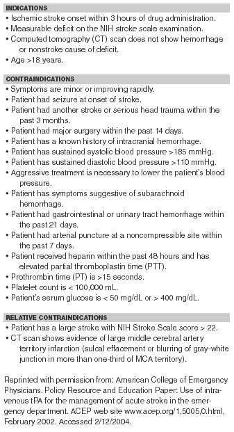
 ACEP - Use of IV tPA for the Management of Acute Stroke in the ED


AHA/ASA Science Advisory - Expansion of the Time Window for Treatment of Acute Ischemic Stroke With Intravenous Tissue Plasminogen Activator (del Zoppo)

ECASS III Exclusion Criteria 
http://crashingpatient.com/medicine-surgery/095-stroke.htm  
Expansion of TPA window to 4.5 hours by AHA (Stroke 2009;40:) - ECASS III
  • Age < 18 or > 80 years
  • Onset of stroke > 4.5 hours before drug administration or symptom onset unknown
  • Stroke symptoms present < 30 minutes or significantly improving before treatment
  • Intracranial hemorrhage
  • Severe stroke as defined by NIHSS > 25 or imaging (CT or MRI) displaying > 1/3 of middle cerebral artery territory involved
  • Seizure at the onset of stroke
  • --------
  • Stroke or serious head trauma within the previous 3-months 
  • Major surgery or severe trauma within 3-months
  • Combination of previous stroke and diabetes mellitus
  • --------
  • SBP > 185 mm Hg or DBP > 110 mm Hg or IV treatment to reduce BP to these limits
  • Glucose < 50 mg/dL or > 400 mg/dL
  • Symptoms suggestive of subarachnoid hemorrhage even if CT normal
  •  -------
  • Heparin within the preceding 48 hours with PTT above normal limit
  • Platelet count < 100,000 mm3
  • Oral anticoagulation therapy (EVEN IF INR WNL)
  • Other major disorders with an increased risk of bleeding


www.aaem.org/education/tpaedtool-AAEM.pdf

Monday, September 19, 2011

Transfusion triggers

Quick note today, just wanted to talk about the hemoglobin level at which we should consider transfusion.

There's a lot of bad data out there, much of it retrospective, heterogeneous populations, and so on. When I was a MICU resident, often the team would prefer to get the Hgb up above 10 to "help with the oxygen delivery." Marino, OTOH, in his The ICU Book, makes a persuasive case that the reduced viscosity in anemia facilitates oxygen delivery, and you mess with this compensatory mechanism (with pRBCs) at your peril. Incidently, Marino provides a review of some more recent transfusion-related papers from his website. Worth reading.

Let me review the single best, and most-oft cited, paper on this topic, which was done by Hêbert et al, published in the NEJM in 1999. The paper (pdf) can be downloaded free from NEJM, I think, but here's a copy.
Real interesting stuff.

They prospectively studied the effect of two different transfusion thresholds, either 7 g/dL (restrictive strategy) or 10 g/dL (liberal strategy), in ICU patients. The ICUs were mixed, both surgical and medical, and both community and academic. They excluded patients with, amongst other issues,  active hemorrhage or s/p cardiac surgery.
They found that 30-day mortality was the same in the two groups, but the hospitalization mortality was significantly lower in the restrictive-strategy group (22.3 percent vs. 28.1 percent, P=0.05).

Let me emphasize that a bit more:
28.1 - 22.3 = 5.8,  or an Absolute Rate Reduction of 5.8%
That gives us a Number Needed to Harm of a little over 17. In other words:

For every 17 patients that were transfused to stay above 10 g/dL, instead of above 7 g/dL, 1 died.


Closer look at the breakdown on adverse effects:
Hey, that's funny. Many folks feel that a chief reason to transfuse is to avoid exacerbating any cardiac ischemia. What they found here, however, was that bad cardiac events were more common with the liberal transfusion threshold!

Okay, that was in 1999 - what's changed since then? Not much. In fact, the evidence has become stronger with regard to the risks of transfusion, and the benefits of a restrictive strategy, across a spectrum of medical and surgical contexts.

Let me give the latest transfusion guidelines, published in 2009, written jointly by the Society for Critical Care Medicine,and the Eastern Association for Surgery on Trauma. This document is kinda unique, addressing both the medical and surgical perspectives regarding the literature. The salient recommendations are copied here. I just want to highlight a few of them:

1. Transfuse patients with active, ongoing hemorrhage. Be proactive!
2. If they're not bleeding out, don't transfuse until the Hgb drops below 7 g/dL.
3. At that point, only transfuse single units.

    A. Recommendations Regarding Indications for RBC Transfusion in the General Critically Ill Patient
      1.    RBC transfusion is indicated for patients with evidence of hemorrhagic shock. (Level 1)  
      2.    RBC transfusion may be indicated for patients with evidence of acute hemorrhage and hemodynamic instability or inadequate oxygen delivery. (Level 1)  
      3.    A “restrictive” strategy of RBC transfusion (transfuse when Hb < 7 g/dL) is as effective as a “liberal” transfusion strategy (transfusion when Hb < 10 g/dL) in critically ill patients with hemodynamically stable anemia, except possibly in patients with acute myocardial ischemia. (Level 1)  
      4.    The use of only Hb level as a “trigger” for transfusion should be avoided. Decision for RBC transfusion should be based on an individual patient's intravascular volume status, evidence of shock, duration and extent of anemia, and cardiopulmonary physiologic parameters. (Level 2) 
      5.    In the absence of acute hemorrhage RBC, transfusion should be given as single units. (Level 2)  
      6.    Consider transfusion if Hb < 7 g/dL in critically ill patients requiring mechanical ventilation (MV). There is no benefit of a “liberal” transfusion strategy (transfusion when Hb < 10 g/dL) in critically ill patients requiring MV. (Level 2)  
      7.    Consider transfusion if Hb < 7 g/dL in resuscitated critically ill trauma patients. There is no benefit of a “liberal” transfusion strategy (transfusion when Hb < 10 g/dL) in resuscitated critically ill trauma patients. (Level 2)  
      8.    Consider transfusion if Hb < 7 g/dL in critically ill patients with stable cardiac disease. There is no benefit of a “liberal” transfusion strategy (transfusion when Hb < 10 g/dL) in critically ill patients with stable cardiac disease. (Level 2) 
      9.    RBC transfusion should not be considered as an absolute method to improve tissue oxygen consumption in critically ill patients. (Level 2) 
      10.  RBC transfusion may be beneficial in patients with acute coronary syndromes (ACS) who are anemic (Hb ≤ 8 g/dL) on hospital admission. (Level 3)
...


Friday, September 16, 2011

Two "facts" about the diagnosis of ACS that you need to forget - Part I

Like the title says, I won't be discussing any new findings or developments in the recognition or diagnosis of ACS that you'll need to memorize, keep on your iPhone, or write on your hand. These are two "important facts" that are hammered into you during your training that are taking up valuable space in your brain, space that could be used for important stuff, like memorizing lines from The Simpsons.

The first thing you need to know is that asking crap about cardiac risk factors is a waste. Sure, it makes the chart look pretty and it gives the appearance of diligence. It also less-than-preferable when you are deep into presenting to an attending, having described in succinct but descriptive language the crushing pain the patient had, the sweating, the left arm heaviness, the relief with nitro, the concerning EKG findings... and all this becomes derailed when the attending asks about family history. "How can you possibly decide what to do with the patient," you are lectured, "if you don't know their risk factors!"

Don't fool yourself into believing that you are obtaining actionable intelligence, however. Knowing whether a patient smokes, takes Lipitor, or had a grampa that keeled over at 45 years of age should not be guiding your diagnostic reasoning.

Okay, so I can avoid sounding like some crazy heretic, let me start off the discussion with an appeal to authority. What do we find in the textbooks about this issue?

In the 2011 Tintinalli, we find that:
“All patients should also be questioned regarding the presence of cardiac risk factors, although these ‘risks’ are valid only for predicting the presence of coronary artery disease within a given population and are not predictive of the presence or absence of acute ischemia in an individual patient.”

Rosen's, as well, comments that:
“The presence of risk factors for a particular disease is primarily of value as an epidemiologic marker… In the ED, presence of risk factors in an individual patient without established disease has minimal or no effect on the clinical likelihood (pretest probability) of a specific disease process.”

Well, is this just the crazy, EM perspective? Nope, here's the American Heart Association in their 2007 NSTEMI guidelines, where they mention the use of risk factors in the initial history:

Even Lee Goldman, a cardiologist renowned for developing various eponymous criteria for determining cardiac risk, has not find the traditional risk factors to be useful, stating that "demographic and traditional risk factors (with the very notable exception of a history of MI or coronary disease) are of little importance in predicting the cause of acute chest pain.”

So, this isn't my wacky opinion, this is actually the conventional wisdom!

Now, perhaps you want some justification, a bit more meat in the argument. Let's check out the empirical evidence in two key studies. The first is the classic systematic review in JAMA, from their excellent Rational Clinical Exam series (results from these papers show up in the in-service!). Written by Panju et al., Is This Patient Having an MI? synthesizes a number of studies looking at the relative contribution of elements of the history, physical exam, and EKG.  There a few shortcomings in the methods, etc., but it is still one of the most oft-cited papers on the topic.
They find that:
Some of this was based on a nice study by Jayes in 1992. Multicenter, prospective collection of information of risk factors in adults being checked out for chest pain. Lotta patients, 1743 in fact. They ran the results through logistic regression and found:
Yeah, there seems to be a small bump in the RR for DM and family history, and barely for smoking. And only in men! These results are blown away by the RR for simply showing up with chest pain, in both men and women.

(Hey, wait a minute. Why is the risk of ACS higher in women with chest pain than for men? Aren't women supposed to present, like, all differently? I'll get to that "fact" in Part II.)

Well, that paper is a few years old now - anything newer out their? Why yes, in fact, there was an excellent single-center study done by Body et al. in Manchester, UK, in the ED. They prospectively obtained information on the standard risk factors (HTN, lipids, DM, smoking, and family history) on 804 adults coming in with "chest pain occurring within the past 24 h that the treating physician suspected to be cardiac in origin." Then they looked at who ruled in/out, and found that knowing the risk factors helped...
... not so much.

Of course, why did we ever expect that these classic risk factors would help us in the ED? This is epidemiological data, used in the office with the asymptomatic patient, to determine the risk of having an event in the next 10 years. Hey, if you're going to bust out your Framingham Risk calculator, knock yourself out, but realize you may be are answering a different question that the one you were aiming for.

You can also listen to this great podcast at ERCAST.

So, when it comes to the utility of cardiac risk factors in the ED, I'm actually not much of a heretic, and I'm not really espousing anything very controversial. I am going out on a bit of a limb, however, in Part II, where I deconstruct the "fact" that women with ACS present with significantly different symptoms and histories than men. Gonna have to show you some evidence there!

Sunday, September 4, 2011

Anaphylactic reactions - 5 things.

I'm a little concerned that the treatment of anaphylaxis is not as well understood by EM folks as it should be. Now, in the acute management of ACS, you can be forgiven for not being familiar about the latest literature regarding the various antithrombotics, the merits of different DES, the proper initial dose of clopidogrel, etc. Too many trials, too much evidence.

But in anaphylaxis. there is hardly any evidence! It's possible to review the entirety of the best evidence in one pithy bog post, so let's get on with it.

First, get this paper. It's the best reference for current diagnosis and treatment of anaphylaxis out there.

Okay, 5 things:

1. Epinephrine is given IM (intramuscular), and in the lateral thigh. Not IM in the deltoid, not SQ in the buttocks. IM in the thigh, period.

The first time I ordered epi this way, the conversation with the nurse went something like this:
"Okay, let's give some epi. I want 0.5 mg IM in the thigh."
"No prob, I already drew it up." (Holds up syringe with 1/2 inch long needle on it)
"Uh, I don't think that'll make it to the muscle..."
"Well, it's only supposed to be SQ, right?"
"No, I want 0.5 mg epi, IM, in the thigh."
"Sure." (attaches longer needle, then rolls up patient's shirt sleeve, exposing the deltoid)
"Hold on a moment - I want that to go in the thigh, not the deltoid."
"You sure? We always do it this way."
"Yes, I want 0.5 mg epi IM in the thigh.
"Okay, you're the doc." (Turns around, eye rolling.)

Just in case you didn't catch it, epinephrine is given IM in the lateral thigh. Quiz later.

Okay, if this is the only thing you learned today, I'm happy. But let's look at the evidence for this, because it's beautiful. Some investigators decided to pay some healthy folks to get epi shots or saline injections. They were administered either IM or SQ, and in the area of either  the deltoid, or lateral thigh. They then measured the serum epi levels found in each of the possible combinations.

So elegant, I could cry. If they didn't give the epi IM in the thigh, the bump in serum epi was no different than placebo. Remember as well, these are healthy volunteers, without any cutaneous vasoconstriction.

2. The dose is 0.01 mg/kg, maxing out at 0.5 mg.  You can repeat this ever 5-15 minutes PRN.

3. The antihistamines and steroids are given only for the (putative) biphasic reaction, and have only a minor role in  acute management. A lot of folks think that this biphasic reaction doesn't even exist, and that "relapses" that occur in the following day or two just represent an incompletely treated MONOphasic reaction. In fact, if you look at this trial that involved giving bee stings to people who had had anaphylactic reactions to bee stings in the past, there is no mention of steroids or antihistamines.

I'm not even bothering to put in the Cochrane Collaboration analyses on the use of these adjunctive measures - both say "No evidence."

4. IV fluids.
They may not have as big a role in trauma these days, but when it comes to anaphylaxis, you want large-bore, proximal IVs, with 2 bags of saline hanging.  A patient in true anaphylactic shock not only has a distributive shock, they actually can lose up to 33% of their intravascular volume in the the initial few minutes. This is the time to break out the 14g catheters!

5.  Intravenous epinephrine
Here  we go - the patient's pressure is tanking, their thigh looks like a pincushion from all the IM epi you've given, and you have the 3rd and 4th liters of saline running in on pressure bags. Time for the IV epi! How do you do this without causing more problems than you already have? Try the 1-1-1 method, probably the best way to give enough, but not too much.

First, grab 1 mg of epinephrine - any concentration will do, the number work out with either the 1:10 or 1:1000 versions.


Next, put this in 1 liter NS.

You'll have a concentration of 1mcg/ml, and you want to start at 1 ml/minute, and titrate up quickly. In the protocol described in the bee sting study above, they would start off at 5 mcg/minute, but then it turns into the 1-1-5 rule. Just titrate up quickly.

Okay, a final word: Isn't epi dangerous? Well, yeah, if you give too much! There are a mess of case reports that show all manner of badness from epinephrine, but most of them appear to result from extraordinary doses.

For example, look at this case: a young adult was given what they describe as "low-dose" IV epi, and subsequently bumped her cardiac enzymes, despite a clean cath. However, what they call "low-dose" was actually 100µg IV, or 20 times the dose that the bee-sting protocol started with, and 100 times what we describe in this post!

Anyway, just remember that you want epinephrine, 0.01mg/kg, given IM, in the lateral thigh!